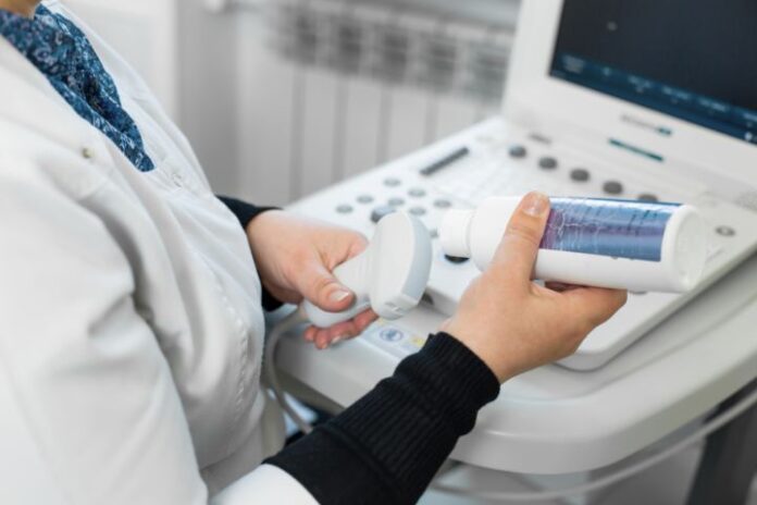
In the realm of veterinary medicine, advancements in technology continue to transform the way veterinarians diagnose and treat ailments in animals. One such groundbreaking technology is veterinary sound wave imaging, commonly known as ultrasound. This non-invasive diagnostic tool has become a cornerstone in modern veterinary practice, offering a safer and more efficient means of understanding and managing pet health issues.
Understanding Veterinary Sound Wave Imaging
Veterinary sound wave imaging utilizes high-frequency sound waves to produce images of the internal structures of an animal’s body. Unlike traditional X-rays, which use radiation, ultrasound imaging is entirely safe and does not expose pets to potentially harmful ionizing radiation. This technology works by sending sound waves into the body using a handheld device called a transducer. The sound waves bounce off internal tissues and organs, creating echoes that are then captured and transformed into real-time images by a computer.
Applications in Veterinary Medicine
The applications of sound wave imaging in veterinary medicine are vast and varied. This technology is used to diagnose and monitor a wide range of conditions, providing veterinarians with crucial insights into the health and well-being of their patients.
- Abdominal Examinations: One of the most common uses of veterinary ultrasound is in the examination of the abdominal cavity. This includes evaluating the liver, kidneys, spleen, bladder, and intestines. Ultrasound can help detect tumors, cysts, and other abnormalities, as well as monitor the progression of diseases such as liver disease or kidney stones.
- Cardiology: Ultrasound imaging plays a pivotal role in veterinary cardiology. It allows for detailed examination of the heart’s structure and function, aiding in the diagnosis of heart diseases such as cardiomyopathy, valvular disease, and congenital heart defects. Echocardiograms, a specific type of ultrasound, provide real-time images of the heart in motion, enabling veterinarians to assess the severity of heart conditions and monitor treatment effectiveness.
- Reproductive Health: In the field of reproductive medicine, ultrasound is indispensable. It is used to confirm pregnancy, monitor fetal development, and detect reproductive issues such as pyometra or ovarian cysts. This non-invasive method is particularly beneficial for breeders and veterinarians working with pregnant animals.
- Musculoskeletal Issues: Ultrasound is also employed to evaluate musculoskeletal problems, including tendon and ligament injuries. It allows for a precise assessment of the extent of injuries, aiding in the development of targeted treatment plans and rehabilitation programs.
Benefits of Veterinary Sound Wave Imaging
The adoption of ultrasound technology in veterinary practice offers numerous benefits:
- Non-Invasive and Painless: Ultrasound is a non-invasive procedure that does not cause pain or discomfort to the animal. This makes it an ideal diagnostic tool, especially for pets that may be anxious or stressed in a clinical setting.
- Real-Time Imaging: One of the significant advantages of ultrasound is its ability to provide real-time images. This allows veterinarians to observe the movement of internal organs, assess blood flow, and make immediate decisions regarding diagnosis and treatment.
- Safe and Radiation-Free: Unlike X-rays, ultrasound does not involve exposure to ionizing radiation, making it a safer option for repeated use, particularly in pregnant animals or those requiring frequent monitoring.
- Comprehensive Diagnostics: Ultrasound offers detailed images of soft tissues, which are often not visible on traditional X-rays. This comprehensive diagnostic capability helps in detecting conditions at an earlier stage, leading to better treatment outcomes.
The Future of Veterinary Sound Wave Imaging
As technology continues to advance, the future of veterinary sound wave imaging looks promising. Innovations such as three-dimensional (3D) and four-dimensional (4D) ultrasound are beginning to make their way into veterinary practice, offering even more detailed and dynamic images. Portable ultrasound devices are also becoming more common, allowing veterinarians to perform diagnostic imaging in various settings, including at-home visits and fieldwork.
Furthermore, the integration of artificial intelligence (AI) into ultrasound technology holds the potential to enhance diagnostic accuracy and efficiency. AI algorithms can assist in interpreting ultrasound images, identifying subtle abnormalities that may be missed by the human eye, and providing veterinarians with valuable insights.
Conclusion
Veterinary sound wave imaging has revolutionized the field of pet diagnostics, providing a safe, non-invasive, and highly effective means of evaluating and monitoring animal health. Its wide range of applications, from abdominal examinations to cardiology and reproductive health, underscores its importance in modern veterinary practice. As technology continues to evolve, the capabilities of ultrasound imaging will only expand, further improving the quality of care provided to our beloved animal companions.


